What if you could saw a wooden sculpture through to look at the tree rings? Could a clay sculpture be hollow? What type of instruments were used to craft art objects? These questions could be answered by X-ray imaging. In particular, CT-scans are useful to investigate historical art objects on the inside. The CT-scans that are suitable for this application are however not easily accessible for researchers and quite expensive to acquire. An international team of researchers led by CWI, the Dutch national research center for mathematics and computer science, has therefore developed a method to use existing X-ray imaging facilities for CT scanning. The results have been published on the 14th of May in Nature Communications.
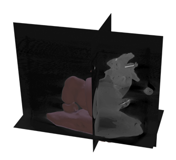
Research facilities of large museums, such as the Rijksmuseum in Amsterdam, often house an X-ray imaging machine, with which they can perform live inspection of objects. This works in the same way as when you have to have an X-ray image taken at the hospital because of a broken bone. On such an image the bone can be seen, because it has a higher density than the muscles and tissue surrounding it. Usually, a few images are taken, so that the fracture can be inspected from different sides. For more complex problems, a CT scan is made. The technique behind CT scans is similar to X-ray images. For a CT scan hundreds of X-ray images are taken from different angles and then combined into a 3D image using reconstruction algorithms. This 3D image can then be ‘sliced open’ to obtain cross sections showing the interior.
Over the past years, this technique has been increasingly applied to art objects, because it gives the possibility to investigate the interior without damaging the object. It is possible to date wooden objects by looking at the tree rings, assess the internal damage by an insect infestation or search for clues about the techniques used by the artist.
No CT scanner? No problem!

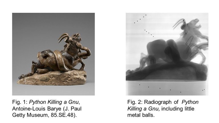
CT scans provide more information than X-ray images. In museums, however, simple X-ray imaging setups are more common. The components of the machine are similar (an X-ray tube, rotation stage and a detector), but for a CT scan the positions of these components during the scan need to be known very accurately. The researchers from CWI have therefore developed a method to calculate these positions after a data acquisition, with nothing more than a few small metal balls. These balls are put in a piece of foam and scanned alongside the object. Based on the location of the balls on the X-ray images, all the necessary positions can be calculated. These are then used to obtain a CT-scan.
This unique project is a collaboration between a team of international researchers from CWI (Amsterdam), Leiden Institute of Advanced Computer Science (Leiden), Rijksmuseum (Amsterdam), The British Museum (London, UK) and the J. Paul Getty Museum (Los Angeles, USA). The X-ray imaging facilities of each of these museums, as well as the FleX-ray laboratory at CWI, have been used to test the new method and compare results in different facilities. The X-ray setups at the Rijksmuseum and the J. Paul Getty museum have been used for CT scans for the first time.
The first author of the associated Nature Communications article Francien Bossema will defend her PhD thesis at Leiden University on the topic of CT scanning for cultural heritage on the 23rd of May.